
Leucine-rich Alpha-2-Glycoprotein-1 and its Binding Partners, Cytochrome c and Transforming Growth Factor-β1, Promote Cancer Progression in an Apoptotic Microenvironment
Professor. Ronald Jemmerson*
Department of Microbiology and Immunology, University of Minnesota, Minneapolis, MN
*Corresponding author: Professor Emeritus. Ronald Jemmerson, Department of Microbiology and Immunology, 689 23rd Ave. S.E., Minneapolis MN 55455, USA; E-mail: jemme001@umn.edu
Running title: LRG1 and its Binding Partners, Cyt c and TGF-β1, Promote Cancer Progression
Received: 27 April 2026; Revised: 18 July 2026; Accepted: 23 July 2026; Published: 27 July 2026
Copyright: © 2026 Jemmerson R. This is an open-access article distributed under the terms of the Creative Commons Attribution License, which permits unrestricted use, distribution, and reproduction in any medium, provided the original author and source are credited.
Citation: Jemmerson R (2026) Leucine-rich Alpha-2-Glycoprotein-1 and its Binding Partners, Cytochrome c and Transforming Growth Factor-β1, Promote Cancer Progression in an Apoptotic Microenvironment. 21st Century Pathology, Volume 6 (1): 162
Abstract
Leucine-rich alpha-2-glycoprotein-1 (LRG1) is an acute-phase protein that increases in circulation when the immune system responds to tissue damage, certain microbial infections, and in many, but not all, cases of cancer. In the last decade, LRG1 has been shown to play roles in the progression of cancer, including cell survival, proliferation, and metastasis. In the apoptotic microenvironment of a tumor, cytochrome c (Cyt c), along with other damage-associated molecular patterns (DAMPs), is released from dying cells. LRG1, also produced by cancer cells, binds Cyt c with high affinity, delays the onset of extracellular Cyt c-induced apoptosis, and blocks its pro-inflammatory effects. Extracellular LRG1 has been shown to signal through the epidermal growth factor (EGF) family of receptors to alter the expression of proteins that regulate the release of Cyt c from mitochondria and, thereby, inhibit apoptosis. Activation of a set of transcription factors via this pathway also promotes proliferation of cancer cells and their migration. Signaling through the transforming growth factor (TGF) receptor on endothelial cells, LRG1 complexed with endoglin and another binding partner, TGF-β1, plays a key role in neovascularization of tumors. This results in dysfunctional and leaky blood vessels, which hinder the delivery of chemotherapeutic drugs to tumors and support cancer cell extravasation to other tissues. TGF-β1 is released from cells of the immune system in response to apoptotic signals, including the exposure of phosphatidylserine (PS) on apoptotic cells and blebs. Potential therapeutic approaches to reduce the effects of LRG1 in pre-clinical testing include a humanized monoclonal antibody (mAb) and a proteolysis targeting chimera (PROTAC) to degrade cytoplasmic LRG1. LRG1 and its binding partners, Cyt c and TGF-β1, provide further insight into the progression of cancer that paradoxically occurs in an apoptotic environment during chemotherapy.
Keywords
Leucine-rich alpha-2-glycoprotein-1 (LRG1); Cytochrome c (Cyt c); Transforming growth factor-b1 (TGF-b1); Cell survival and Apoptotic microenvironment
Introduction
Conceptualizations of the causes of cancer have evolved over the years from focusing on alterations within the cells to inclusion of factors in the microenvironment such as those emanating from apoptotic cells [1-5]. Paradoxically, chemotherapy, which induces apoptosis, can result in more aggressive forms of cancer despite the intent to kill cancer cells [4,5]. Some of the factors released from or exposed on apoptotic cells that play a role in cancer progression have been identified [4,5]. For example, ATP and other metabolites can be taken up into cancer cells, providing energy and nutrients for growth and survival and suppression of inflammation [6]. Prostaglandin E2, derived downstream following the activation of calcium-independent phospholipase A2 by caspases, stimulates cancer cell proliferation [7,8]. Phosphatidylserine (PS), which is exposed on the outer leaflet of apoptotic cells and blebs released from the cells, induces endothelial cell sprouting leading to aberrant angiogenesis in tumors [9].
LRG1 (leucine-rich alpha-2-glycoprotein-1) and two of its binding partners, cytochrome c (Cyt c) and transforming growth factor-β1 (TGF-β1), are, potentially, additional key players in cancer cell progression in an apoptotic microenvironment. LRG1 plays a key role in aberrant angiogenesis in tumors and has pro-survival, proliferative, and metastatic effects on many types of cancer cells [10,11]. LRG1 is a ubiquitous biological constituent whose normal role may be in maintaining tissue homeostasis under stress conditions by limiting tissue damage from infection or other injury [12]. In an aberrant extension of this function, LRG1 also impacts the progression of cancer [10,11].
LRG1 and its Binding Partners, Cyt c and TGF-β1
LRG1 was first isolated from human serum in 1977 [13]. It is a 50 kD glycoprotein produced largely by the liver, but also by neutrophils and other cells [14]. LRG1 is characterized as an acute-phase protein produced in response to tissue damage and infection by some microbes [15]. Increased serum levels of LRG1 occur in many diseases and often is prognostic in cancer, indicating a poor outcome [10,11,16]. The amino acid sequence of LRG1 was determined in 1985, revealing repetitive leucine-rich sequences. It is the first identified member of what is now known as the very large family of proteins containing leucine-rich repeats [17]. LRG1 assumes a horseshoe shape that is typical of proteins with this repetitive motif (Figure 1) [18]. Interactions of members of this family with other proteins generally occur along the concave surface [18,19].
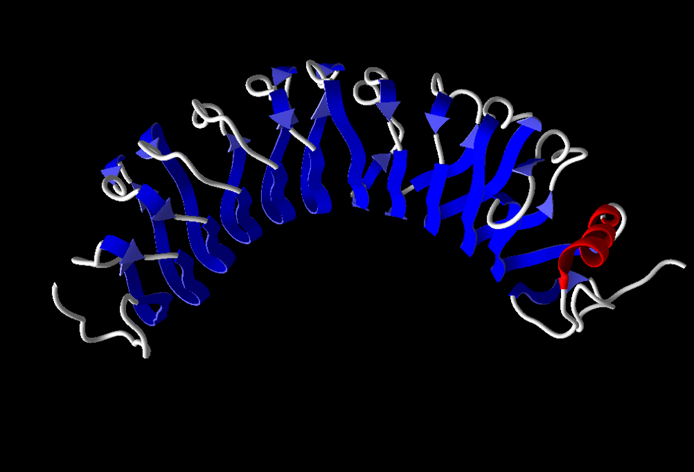
Figure 1: The molecular model of LRG1 obtained employing the computer program Molegro (University of Illinois, Urbana-Champaign) is very similar to the structure that was determined by X-ray crystallography (18).
Twenty-five years after the discovery of LRG1, clues of its function(s) began to emerge with the identification of binding partners. In 2002, TGF-β1 was shown to bind a previously unknown protein in mice that is a marker for endothelial cells and was later identified as the mouse homolog of human LRG1 [20]. In 2006, Cyt c was discovered as a ligand when LRG1 was found to be the component in serum that interfered with detection of Cyt c in an antibody-based sandwich enzyme-linked immunosorbent assay [21]. A molecular model of Cyt c bound to LRG1 has been proposed (see Supplementary Material in ref. 18). In this model, Cyt c, which is positively charged, occupies the space beneath the negatively charged concave surface shown in Figure 1. A model for TGF-β1 bound to LRG1 has not been published. Binding of these ligands to LRG1 has been confirmed employing surface plasmon resonance and the affinities determined. LRG1 has an affinity (Kd) for Cyt c of 1.58 x 10⁻¹³ M and a much lower affinity for TGF-β1 of 2.32 x 10⁻⁶ M [20,22].
Pro-apoptotic Effect of Extracellular Cyt c
In apoptosis, Cyt c translocates from mitochondria into the cytoplasm, where it binds Apaf-1, leading to the activation of caspase-9, initiating an enzymatic (caspase) cascade that culminates in cell death [23,24]. Eventually, Cyt c is released from apoptotic cells in vitroand in vivo if apoptotic bodies are not effectively engulfed by phagocytic cells [25-27]. The protein has been detected in the blood of healthy individuals and is elevated in the blood of cancer patients receiving chemotherapy [27]. Cyt c is among a cohort of molecules released from dying cells that are referred to as DAMPs (damage-associated molecular patterns) [28]. As an extracellular protein, Cyt c, in amounts found to be released from apoptotic cells in vitro, has been shown to enhance apoptosis when added to cultures of neurons or lymphocytes [26,29]. The increased apoptosis was blocked by the addition of Cyt c-specific antibodies, confirming the role of extracellular Cyt c in the induction of apoptosis [26,29]. The pro-apoptotic effect of extracellular Cyt c has also been observed with the cancer cell line J774, which is derived from a mouse reticulosarcoma, showing that the toxic effect of Cyt c is not limited to normal cells [30].
Pro-survival Function of LRG1 Interacting with Cyt
Considering the evidence that extracellular Cyt c is pro-apoptotic and that LRG1 binds Cyt c, a function of LRG1 could be to protect cells in an apoptotic environment from Cyt c-induced toxicity [29]. In 2010 it was reported that the addition of LRG1 to LRG1-depleted medium supplemented with Cyt c extended survival of both human and mouse lymphocytes by several days. LRG1 also extended the life of lymphocytes without adding Cyt c to the cultures because cells began to release their own Cyt c as nutrients in the medium were depleted [29].
Indirect evidence that was obtained supports the idea that the complex of LRG1 and Cyt c delivers a survival signal [29]. The most convincing evidence for a signalling complex is that LRG1 at an optimum concentration of 2 x 10⁻⁸ M delays apoptosis induced by Cyt c at a concentration of 2 x 10⁻⁶ M. The vast molar excess of Cyt c compared to LRG1 indicates that LRG1 does not simply sterically block Cyt c. Also, in response to the addition of horse Cyt c to the cultures, human lymphocytes were protected slightly more effectively by human LRG1 than by mouse LRG1 and mouse lymphocytes were protected more effectively by mouse LRG1 than human LRG1 suggesting the existence of a species-specific receptor. Finally, LRG1 must be bound to Cyt c and does not deliver a survival signal independently of Cyt c as LRG1 did not protect lymphocytes from the toxicity of the tri-methyllysine 72 variant of Cyt c that LRG1 fails to bind [29].
LRG1 is Immunosuppressive and Blocks the Pro-inflammatory Effect of Cyt c
In 2016 LRG1 was shown to play a role in inflammation in a mouse model of myocardial infarction induced by occlusion of a coronary artery [31]. Mice in which the lrg1 gene had been knocked out developed a more severe case of fibrosis resulting from chronic inflammation. Transplantation of myeloid cells into these mice, including neutrophils which produce LRG1, attenuated fibrosis [31].
More recently, a study in obese mice showed that LRG1 inhibited macrophage production of pro-inflammatory cytokines induced by Cyt c in vitro[32]. LRG1 had been depleted from the culture medium and then added back to demonstrate inhibition by LRG1. When LRG1 was elevated in mice fed a high-fat diet employing a viral vector containing the lrg1 gene, inflammation was suppressed. Complexes of LRG1 and Cyt c were observed in the blood, with increased levels in mice fed a high-fat diet compared to mice fed a low-fat diet [32].
In earlier studies, Cyt c had been shown to induce the production of pro-inflammatory cytokines including TNF-a and IL-6 by mouse spleen cells and GM-CSF and IL-1b by human astrocytes [33,34]. In mice, Cyt c injected into the knee joints induced transient arthritis lasting less than 10 days [33]. In the study of astrocytes, an antibody against Toll-like receptor 4 blocked the effect of Cyt c suggesting that it was the receptor transmitting the signal leading to cytokine production [34].
Angiogenic Function of LRG1 Interacting with TGF-β1
TGF-β1 is ubiquitous and is also produced during tissue injury and repair by cells of the immune system [35]. Apoptotic cells in this environment activate macrophages to secrete TGF-β1 in response to PS exposure on their outer membrane [36]. The complex of LRG1, TGF-β1, and endoglin was shown in 2013 to play a key role in neovascularization in eye disease by signalling through the canonical TbRII pathway, in association with ALK1 [37]. This complex has since been shown to play a key role in tumor angiogenesis (neovascularization). The newly formed vessels are often leaky and weakened, which negatively impacts drug delivery and facilitates extravasation of cancer cells to other tissues [38].
LRG1 Promotes Cancer Cell Survival, Proliferation, and Migration
In 2015, early evidence that LRG1 promotes cancer cell survival was obtained by decreasing the expression of LRG1 in cultured glioblastoma cells employing short-hairpin RNA (shRNA) to target LRG1 expression [39]. The treated cells were decreased in number, and some of them assumed an apoptotic phenotype, i.e., binding to annexin V, which indicates PS exposure on the outer membrane. By Western blotting, there was a 2 to 3-fold increase in the pro-apoptotic protein Bax, which promotes the release of Cyt c from mitochondria, a 2- to 3-fold decrease in the anti-apoptotic protein Bcl-2, which blocks the release of Cyt c, along with decreased expression of cyclins D1, B, and E, indicating cell cycle arrest. in vivo inhibition of tumor growth in mice by more than 2-fold was also demonstrated for glioblastoma cells treated with shRNA targeting the expression of LRG1 [39].
These results have been confirmed in several reports employing similar approaches, including studies of colorectal cancer, pancreatic cancer, and multiple myeloma [40-42]. In three independent studies of colorectal and pancreatic cancers, EGF family receptors (EGFR or HER1 and HER3) were shown to be involved in LRG1 signaling indicating a separate pathway from that leading to neovascularization which involves TbRII [41,43,44].
In addition to demonstrating a role for LRG1 in cancer cell survival and proliferation, multiple studies also showed its role in cancer cell migration by the effect of LRG1 on expression of cell-surface adhesion molecules N-cadherin and E-cadherin [45-47]. The level of LRG1 was decreased using RNA silencing to show that E-cadherin was increased and N-caderin was decreased in cells, while overexpression of LRG1 by gene transfection resulted in opposite levels of these two transmembrane glycoproteins. An increase in N-cadherin and decrease in E-cadherin are consistent with the epithelial-to-mesenchymal transition (EMT), which is a hallmark of metastasis [48].
The 2015 study of glioblastoma cells did not determine whether the survival effect was due to extracellular LRG1 or intracellular LRG1 [39]. In an in vitrostudy of colorectal cancer cells in which LRG1 was also decreased using shRNA, recombinant LRG1 added to the cultures prolonged cell survival and reversed apoptosis [40]. In more recent experiments by others, colorectal cancer cells were introduced into lrg1 gene knockout mice and cancer cell proliferation was compared to normal mice [43]. Tumor growth was suppressed in mice lacking LRG1. In addition, an anti-LRG1 mAb lessened tumor burden when cancer cells were transplanted into normal mice [43]. These studies clearly demonstrated that extracellular LRG1 promoted cell survival. LRG1 is often overexpressed in cancer cells and secreted as an autocrine to exert extracellular effects, in addition to the amounts released into blood from normal cells such as hepatocytes and neutrophils [14,40,43].
Schematic Summary
Figure 2 represents a schematic summary of the key discussion points. On the left in the figure is depicted a vascularized tumor infiltrated with cells of the immune system. For simplicity, only lymphocytes and macrophages are shown. As a result of chemotherapy, cells begin to undergo apoptosis, as depicted in the middle section. These include not only cancer cells but also cells of the immune system.
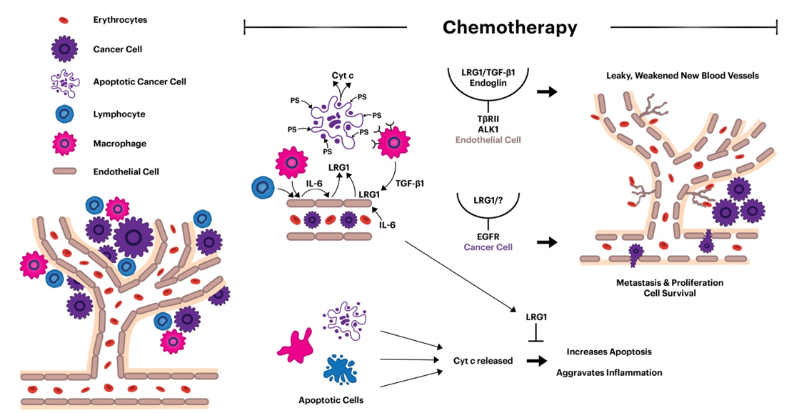
Figure 2: Schematic cartoon showing highlights of the effects of LRG1 and its ligands, Cyt c and TGF-β1, in an apoptotic microenvironment. See the text for details.
In the middle of the figure at the bottom, Cyt c and other DAMPs are released into the microenvironment. The amounts that accumulate depend on the efficiency of phagocytosis. The released Cyt c can induce apoptosis from the outside in cells not affected by the chemotherapy and aggravate inflammation by inducing macrophages to release pro-inflammatory cytokines. Cyt c, by itself, should be helpful in eliminating cancer cells. However, LRG1 blocks these functions of Cyt c.
In the middle of the figure at the top, cells of the immune system and endothelial cells lining the blood vessels are shown to produce IL-6 which induces the production of LRG1 by endothelial cells. Neutrophils and cancer cells overexpressing LRG1 also secrete the glycoprotein. Receptors on macrophages bind PS and then secrete TGF-β1 in response. The complex of TGF-β1/LRG1 and endoglin on endothelial cells activates the canonical TGF-β signalling pathway in aberrant neovascularization resulting in leaky and weakened blood vessels. This prevents effective delivery of chemotherapeutic drugs and enhances the ability of the tumor cells to extravasate. Interaction of LRG1 with EGFR family members on the cancer cell surface promotes cancer progression by transmitting anti-apoptotic, proliferative, and pro-metastatic signals affecting gene transcription. The “?” in conjunction with LRG1 represents a potential ligand, possibly Cyt c.
Potential Therapies
A mouse mAb that blocks neovascularization of tumors induced by LRG1and normalizes the vasculature has been effective in improving the delivery of chemotherapeutic drugs to mouse tumors [38]. The mAb alone blocked the anti-apoptotic, proliferative, and metastatic functions of LRG1, reducing tumor volume in mice by 39% and, in combination with cisplatin, reducing tumor volume by 25% more than cisplatin alone. The anti-LRG1 mAb also enhanced the immune checkpoint blockade effect of mAb anti-PD-1, reducing tumor volume by 88%, a statistically significant improvement over anti-PD-1 treatment alone [38]. Pre-clinical testing of a humanized version of the antibody is underway in the United Kingdom [49,50]. More than 40 mAbs are currently in clinical use for cancer therapy [51].
A proteolysis targeting chimera (PROTAC) has been developed to deliver cytoplasmic LRG1 to proteasomes for degradation [52]. By reducing intracellular LRG1, less is secreted to signal through cell-surface receptors and intracellular functions that inhibit apoptosis and promote proliferation, and metastasis is blocked. The PROTAC contains a short LRG1-binding peptide linked to a molecule that binds a ligand of the E3 ubiquitin ligase. This molecular glue allows ubiquitination of LRG1, directing its degradation in proteasomes. The LRG1-specific PROTAC has been modified into a nanoparticle that allows it to escape the acidic environment of the endosome and enter the cytoplasm. In mice, this nano-PROTAC was preferentially located in tumors, although also in the liver to a lesser extent, and reduced tumor size 3 to 4-fold. No side effects have been observed in mice [52]. At least 30 PROTACs targeting different molecules are in various stages of clinical trials, although none have yet been approved for cancer therapy [53].
Enhancing phagocytic activity could minimize the effect of apoptotic cells in cancer progression. Research in this area is focused on understanding the surface molecules that prompt phagocytic cells to engulf apoptotic cells and to exploit that knowledge to more efficiently clear apoptotic debris [54]. In a recent report, LRG1 was shown to decrease phagocytosis of neutrophils by microglial cells [55]. This may infer that another role for LRG1 in promoting cancer progression in an apoptotic microenvironment is to inhibit phagocytosis of apoptotic cells. This invites further investigation.
Caveat Regarding the Association of LRG1 with Cancer
While LRG1 is generally elevated in the blood of cancer patients, predicting a poor outcome, for prostate cancer patients, elevated levels of LRG1 predict reduced risk of recurrence and castration-resistant cancer [56]. There are cancer cell lines expressing lower levels of LRG1. In these cells, LRG1 has been shown to be pro-apoptotic [57,58]. The distinction from other cancer cells appears to be LRG1 signalling through a different receptor (TbRI and not TbRII, nor EGFR/HER3). This indicates different internal chemical connections leading to expression of a distinct set of genes.
Conclusions
Understanding the factors associated with apoptotic cells that drive the progression of cancer may lead to alternative or supplementary approaches to cancer therapy. LRG1 and its ligands should be added to the list as key players. LRG1 is often present at higher levels in the blood of cancer patients and is a prognostic indicator of a poor outcome. One of the LRG1 binding partners, Cyt c, is released from apoptotic cancer and other cells in response to chemotherapy and can induce apoptosis in neighbouring cells. LRG1 blocks this effect of Cyt c, perhaps by delivering a pro-survival signal to the cells. LRG1 is also immunosuppressive by inhibiting the pro-inflammatory effect of Cyt c. It has been proposed that the normal function of LRG1 may be to act as a sensor detecting Cyt c released from dying cells and then, in complex with Cyt c, to deliver a signal protecting cells in the local environment from the pro-apoptotic effect of Cyt c and, perhaps, other DAMPS. Unfortunately, this putative homeostasis function of LRG1 is also available for survival of cancer cells. Another binding partner of LRG1, TGF-β1, is secreted by immune cells infiltrating the apoptotic environment. Together, LRG1 and TGF-β1 play a key role in the aberrant neovascularization of tumors resulting in weakened and leaky blood vessels that facilitate extravasation of cancer cells and hinder the effective delivery of chemotherapeutic drugs. Anti-LRG1 mAbs have been employed successfully in mice to inhibit the proliferation and metastasis of cancer cells as well as to normalize tumor vasculature. A humanized version of one of the mAbs is in pre-clinical testing in the United Kingdom, giving hope for a novel target in anti-cancer therapy. In addition, a PROTAC degrading cytoplasmic LRG1 has been developed that reduces the amount of LRG1 secreted to act as an autocrine, blocks its anti-apoptotic function, and successfully diminishes the size of tumors in mice. This potential therapy is still in the experimental stage.
List of Abbreviations
Cyt c: Cytochrome c; DAMPs: Damage-associated molecular patterns; EGFR or HER1: Epidermal growth factor receptor or human epidermal growth factor receptor 1; HER3: Human epidermal growth factor receptor 3; IL-6: Interleukin-6; LRG1: Leucine-rich alpha-2-glycoprotein-1; mAb: Monoclonal antibody; PROTAC: Proteolysis targeting chimera; PS: Phosphatidylserine; TbRII or TbRI: Receptors for TGF-β1; TGF-β1: Transforming growth factor-β1; shRNA: Short-hairpin RNA
Acknowledgments
This publication was not supported by external funds.
Conflict of Interest Statement
The author has no competing interests.
Author Contributions
Conceptualization, drafting, editing, and final approval: RJ; computer graphics design: RJ (computer graphics implementation: Katie Leonardis)
Institutional Review Board
Not applicable (no new reporting of human or animal research)
Introduction
Conceptualizations of the causes of cancer have evolved over the years from focusing on alterations within the cells to inclusion of factors in the microenvironment such as those emanating from apoptotic cells [1-5]. Paradoxically, chemotherapy, which induces apoptosis, can result in more aggressive forms of cancer despite the intent to kill cancer cells [4,5]. Some of the factors released from or exposed on apoptotic cells that play a role in cancer progression have been identified [4,5]. For example, ATP and other metabolites can be taken up into cancer cells, providing energy and nutrients for growth and survival and suppression of inflammation [6]. Prostaglandin E2, derived downstream following the activation of calcium-independent phospholipase A2 by caspases, stimulates cancer cell proliferation [7,8]. Phosphatidylserine (PS), which is exposed on the outer leaflet of apoptotic cells and blebs released from the cells, induces endothelial cell sprouting leading to aberrant angiogenesis in tumors [9].
LRG1 (leucine-rich alpha-2-glycoprotein-1) and two of its binding partners, cytochrome c (Cyt c) and transforming growth factor-β1 (TGF-β1), are, potentially, additional key players in cancer cell progression in an apoptotic microenvironment. LRG1 plays a key role in aberrant angiogenesis in tumors and has pro-survival, proliferative, and metastatic effects on many types of cancer cells [10,11]. LRG1 is a ubiquitous biological constituent whose normal role may be in maintaining tissue homeostasis under stress conditions by limiting tissue damage from infection or other injury [12]. In an aberrant extension of this function, LRG1 also impacts the progression of cancer [10,11].
LRG1 and its Binding Partners, Cyt c and TGF-β1
LRG1 was first isolated from human serum in 1977 [13]. It is a 50 kD glycoprotein produced largely by the liver, but also by neutrophils and other cells [14]. LRG1 is characterized as an acute-phase protein produced in response to tissue damage and infection by some microbes [15]. Increased serum levels of LRG1 occur in many diseases and often is prognostic in cancer, indicating a poor outcome [10,11,16]. The amino acid sequence of LRG1 was determined in 1985, revealing repetitive leucine-rich sequences. It is the first identified member of what is now known as the very large family of proteins containing leucine-rich repeats [17]. LRG1 assumes a horseshoe shape that is typical of proteins with this repetitive motif (Figure 1) [18]. Interactions of members of this family with other proteins generally occur along the concave surface [18,19].
Figure 1: The molecular model of LRG1 obtained employing the computer program Molegro (University of Illinois, Urbana-Champaign) is very similar to the structure that was determined by X-ray crystallography (18).
Twenty-five years after the discovery of LRG1, clues of its function(s) began to emerge with the identification of binding partners. In 2002, TGF-β1 was shown to bind a previously unknown protein in mice that is a marker for endothelial cells and was later identified as the mouse homolog of human LRG1 [20]. In 2006, Cyt c was discovered as a ligand when LRG1 was found to be the component in serum that interfered with detection of Cyt c in an antibody-based sandwich enzyme-linked immunosorbent assay [21]. A molecular model of Cyt c bound to LRG1 has been proposed (see Supplementary Material in ref. 18). In this model, Cyt c, which is positively charged, occupies the space beneath the negatively charged concave surface shown in Figure 1. A model for TGF-β1 bound to LRG1 has not been published. Binding of these ligands to LRG1 has been confirmed employing surface plasmon resonance and the affinities determined. LRG1 has an affinity (Kd) for Cyt c of 1.58 x 10⁻¹³ M and a much lower affinity for TGF-β1 of 2.32 x 10⁻⁶ M [20,22].
Pro-apoptotic Effect of Extracellular Cyt c
In apoptosis, Cyt c translocates from mitochondria into the cytoplasm, where it binds Apaf-1, leading to the activation of caspase-9, initiating an enzymatic (caspase) cascade that culminates in cell death [23,24]. Eventually, Cyt c is released from apoptotic cells in vitro and in vivo if apoptotic bodies are not effectively engulfed by phagocytic cells [25-27]. The protein has been detected in the blood of healthy individuals and is elevated in the blood of cancer patients receiving chemotherapy [27]. Cyt c is among a cohort of molecules released from dying cells that are referred to as DAMPs (damage-associated molecular patterns) [28]. As an extracellular protein, Cyt c, in amounts found to be released from apoptotic cells in vitro, has been shown to enhance apoptosis when added to cultures of neurons or lymphocytes [26,29]. The increased apoptosis was blocked by the addition of Cyt c-specific antibodies, confirming the role of extracellular Cyt c in the induction of apoptosis [26,29]. The pro-apoptotic effect of extracellular Cyt c has also been observed with the cancer cell line J774, which is derived from a mouse reticulosarcoma, showing that the toxic effect of Cyt c is not limited to normal cells [30].
Pro-survival Function of LRG1 Interacting with Cyt
Considering the evidence that extracellular Cyt c is pro-apoptotic and that LRG1 binds Cyt c, a function of LRG1 could be to protect cells in an apoptotic environment from Cyt c-induced toxicity [29]. In 2010 it was reported that the addition of LRG1 to LRG1-depleted medium supplemented with Cyt c extended survival of both human and mouse lymphocytes by several days. LRG1 also extended the life of lymphocytes without adding Cyt c to the cultures because cells began to release their own Cyt c as nutrients in the medium were depleted [29].
Indirect evidence that was obtained supports the idea that the complex of LRG1 and Cyt c delivers a survival signal [29]. The most convincing evidence for a signalling complex is that LRG1 at an optimum concentration of 2 x 10⁻⁸ M delays apoptosis induced by Cyt c at a concentration of 2 x 10⁻⁶ M. The vast molar excess of Cyt c compared to LRG1 indicates that LRG1 does not simply sterically block Cyt c. Also, in response to the addition of horse Cyt c to the cultures, human lymphocytes were protected slightly more effectively by human LRG1 than by mouse LRG1 and mouse lymphocytes were protected more effectively by mouse LRG1 than human LRG1 suggesting the existence of a species-specific receptor. Finally, LRG1 must be bound to Cyt c and does not deliver a survival signal independently of Cyt c as LRG1 did not protect lymphocytes from the toxicity of the tri-methyllysine 72 variant of Cyt c that LRG1 fails to bind [29].
LRG1 is Immunosuppressive and Blocks the Pro-inflammatory Effect of Cyt c
In 2016 LRG1 was shown to play a role in inflammation in a mouse model of myocardial infarction induced by occlusion of a coronary artery [31]. Mice in which the lrg1 gene had been knocked out developed a more severe case of fibrosis resulting from chronic inflammation. Transplantation of myeloid cells into these mice, including neutrophils which produce LRG1, attenuated fibrosis [31].
More recently, a study in obese mice showed that LRG1 inhibited macrophage production of pro-inflammatory cytokines induced by Cyt c in vitro[32]. LRG1 had been depleted from the culture medium and then added back to demonstrate inhibition by LRG1. When LRG1 was elevated in mice fed a high-fat diet employing a viral vector containing the lrg1 gene, inflammation was suppressed. Complexes of LRG1 and Cyt c were observed in the blood, with increased levels in mice fed a high-fat diet compared to mice fed a low-fat diet [32].
In earlier studies, Cyt c had been shown to induce the production of pro-inflammatory cytokines including TNF-a and IL-6 by mouse spleen cells and GM-CSF and IL-1b by human astrocytes [33,34]. In mice, Cyt c injected into the knee joints induced transient arthritis lasting less than 10 days [33]. In the study of astrocytes, an antibody against Toll-like receptor 4 blocked the effect of Cyt c suggesting that it was the receptor transmitting the signal leading to cytokine production [34].
Angiogenic Function of LRG1 Interacting with TGF-β1
TGF-β1 is ubiquitous and is also produced during tissue injury and repair by cells of the immune system [35]. Apoptotic cells in this environment activate macrophages to secrete TGF-β1 in response to PS exposure on their outer membrane [36]. The complex of LRG1, TGF-β1, and endoglin was shown in 2013 to play a key role in neovascularization in eye disease by signalling through the canonical TbRII pathway, in association with ALK1 [37]. This complex has since been shown to play a key role in tumor angiogenesis (neovascularization). The newly formed vessels are often leaky and weakened, which negatively impacts drug delivery and facilitates extravasation of cancer cells to other tissues [38].
LRG1 Promotes Cancer Cell Survival, Proliferation, and Migration
In 2015, early evidence that LRG1 promotes cancer cell survival was obtained by decreasing the expression of LRG1 in cultured glioblastoma cells employing short-hairpin RNA (shRNA) to target LRG1 expression [39]. The treated cells were decreased in number, and some of them assumed an apoptotic phenotype, i.e., binding to annexin V, which indicates PS exposure on the outer membrane. By Western blotting, there was a 2 to 3-fold increase in the pro-apoptotic protein Bax, which promotes the release of Cyt c from mitochondria, a 2- to 3-fold decrease in the anti-apoptotic protein Bcl-2, which blocks the release of Cyt c, along with decreased expression of cyclins D1, B, and E, indicating cell cycle arrest. in vivo inhibition of tumor growth in mice by more than 2-fold was also demonstrated for glioblastoma cells treated with shRNA targeting the expression of LRG1 [39].
These results have been confirmed in several reports employing similar approaches, including studies of colorectal cancer, pancreatic cancer, and multiple myeloma [40-42]. In three independent studies of colorectal and pancreatic cancers, EGF family receptors (EGFR or HER1 and HER3) were shown to be involved in LRG1 signaling indicating a separate pathway from that leading to neovascularization which involves TbRII [41,43,44].
In addition to demonstrating a role for LRG1 in cancer cell survival and proliferation, multiple studies also showed its role in cancer cell migration by the effect of LRG1 on expression of cell-surface adhesion molecules N-cadherin and E-cadherin [45-47]. The level of LRG1 was decreased using RNA silencing to show that E-cadherin was increased and N-caderin was decreased in cells, while overexpression of LRG1 by gene transfection resulted in opposite levels of these two transmembrane glycoproteins. An increase in N-cadherin and decrease in E-cadherin are consistent with the epithelial-to-mesenchymal transition (EMT), which is a hallmark of metastasis [48].
The 2015 study of glioblastoma cells did not determine whether the survival effect was due to extracellular LRG1 or intracellular LRG1 [39]. In an in vitrostudy of colorectal cancer cells in which LRG1 was also decreased using shRNA, recombinant LRG1 added to the cultures prolonged cell survival and reversed apoptosis [40]. In more recent experiments by others, colorectal cancer cells were introduced into lrg1 gene knockout mice and cancer cell proliferation was compared to normal mice [43]. Tumor growth was suppressed in mice lacking LRG1. In addition, an anti-LRG1 mAb lessened tumor burden when cancer cells were transplanted into normal mice [43]. These studies clearly demonstrated that extracellular LRG1 promoted cell survival. LRG1 is often overexpressed in cancer cells and secreted as an autocrine to exert extracellular effects, in addition to the amounts released into blood from normal cells such as hepatocytes and neutrophils [14,40,43].
Schematic Summary
Figure 2 represents a schematic summary of the key discussion points. On the left in the figure is depicted a vascularized tumor infiltrated with cells of the immune system. For simplicity, only lymphocytes and macrophages are shown. As a result of chemotherapy, cells begin to undergo apoptosis, as depicted in the middle section. These include not only cancer cells but also cells of the immune system.
Figure 2: Schematic cartoon showing highlights of the effects of LRG1 and its ligands, Cyt c and TGF-β1, in an apoptotic microenvironment. See the text for details.
In the middle of the figure at the bottom, Cyt c and other DAMPs are released into the microenvironment. The amounts that accumulate depend on the efficiency of phagocytosis. The released Cyt c can induce apoptosis from the outside in cells not affected by the chemotherapy and aggravate inflammation by inducing macrophages to release pro-inflammatory cytokines. Cyt c, by itself, should be helpful in eliminating cancer cells. However, LRG1 blocks these functions of Cyt c.
In the middle of the figure at the top, cells of the immune system and endothelial cells lining the blood vessels are shown to produce IL-6 which induces the production of LRG1 by endothelial cells. Neutrophils and cancer cells overexpressing LRG1 also secrete the glycoprotein. Receptors on macrophages bind PS and then secrete TGF-β1 in response. The complex of TGF-β1/LRG1 and endoglin on endothelial cells activates the canonical TGF-β signalling pathway in aberrant neovascularization resulting in leaky and weakened blood vessels. This prevents effective delivery of chemotherapeutic drugs and enhances the ability of the tumor cells to extravasate. Interaction of LRG1 with EGFR family members on the cancer cell surface promotes cancer progression by transmitting anti-apoptotic, proliferative, and pro-metastatic signals affecting gene transcription. The “?” in conjunction with LRG1 represents a potential ligand, possibly Cyt c.
Potential Therapies
A mouse mAb that blocks neovascularization of tumors induced by LRG1and normalizes the vasculature has been effective in improving the delivery of chemotherapeutic drugs to mouse tumors [38]. The mAb alone blocked the anti-apoptotic, proliferative, and metastatic functions of LRG1, reducing tumor volume in mice by 39% and, in combination with cisplatin, reducing tumor volume by 25% more than cisplatin alone. The anti-LRG1 mAb also enhanced the immune checkpoint blockade effect of mAb anti-PD-1, reducing tumor volume by 88%, a statistically significant improvement over anti-PD-1 treatment alone [38]. Pre-clinical testing of a humanized version of the antibody is underway in the United Kingdom [49,50]. More than 40 mAbs are currently in clinical use for cancer therapy [51].
A proteolysis targeting chimera (PROTAC) has been developed to deliver cytoplasmic LRG1 to proteasomes for degradation [52]. By reducing intracellular LRG1, less is secreted to signal through cell-surface receptors and intracellular functions that inhibit apoptosis and promote proliferation, and metastasis is blocked. The PROTAC contains a short LRG1-binding peptide linked to a molecule that binds a ligand of the E3 ubiquitin ligase. This molecular glue allows ubiquitination of LRG1, directing its degradation in proteasomes. The LRG1-specific PROTAC has been modified into a nanoparticle that allows it to escape the acidic environment of the endosome and enter the cytoplasm. In mice, this nano-PROTAC was preferentially located in tumors, although also in the liver to a lesser extent, and reduced tumor size 3 to 4-fold. No side effects have been observed in mice [52]. At least 30 PROTACs targeting different molecules are in various stages of clinical trials, although none have yet been approved for cancer therapy [53].
Enhancing phagocytic activity could minimize the effect of apoptotic cells in cancer progression. Research in this area is focused on understanding the surface molecules that prompt phagocytic cells to engulf apoptotic cells and to exploit that knowledge to more efficiently clear apoptotic debris [54]. In a recent report, LRG1 was shown to decrease phagocytosis of neutrophils by microglial cells [55]. This may infer that another role for LRG1 in promoting cancer progression in an apoptotic microenvironment is to inhibit phagocytosis of apoptotic cells. This invites further investigation.
Caveat Regarding the Association of LRG1 with Cancer
While LRG1 is generally elevated in the blood of cancer patients, predicting a poor outcome, for prostate cancer patients, elevated levels of LRG1 predict reduced risk of recurrence and castration-resistant cancer [56]. There are cancer cell lines expressing lower levels of LRG1. In these cells, LRG1 has been shown to be pro-apoptotic [57,58]. The distinction from other cancer cells appears to be LRG1 signalling through a different receptor (TbRI and not TbRII, nor EGFR/HER3). This indicates different internal chemical connections leading to expression of a distinct set of genes.
Conclusions
Understanding the factors associated with apoptotic cells that drive the progression of cancer may lead to alternative or supplementary approaches to cancer therapy. LRG1 and its ligands should be added to the list as key players. LRG1 is often present at higher levels in the blood of cancer patients and is a prognostic indicator of a poor outcome. One of the LRG1 binding partners, Cyt c, is released from apoptotic cancer and other cells in response to chemotherapy and can induce apoptosis in neighbouring cells. LRG1 blocks this effect of Cyt c, perhaps by delivering a pro-survival signal to the cells. LRG1 is also immunosuppressive by inhibiting the pro-inflammatory effect of Cyt c. It has been proposed that the normal function of LRG1 may be to act as a sensor detecting Cyt c released from dying cells and then, in complex with Cyt c, to deliver a signal protecting cells in the local environment from the pro-apoptotic effect of Cyt c and, perhaps, other DAMPS. Unfortunately, this putative homeostasis function of LRG1 is also available for survival of cancer cells. Another binding partner of LRG1, TGF-β1, is secreted by immune cells infiltrating the apoptotic environment. Together, LRG1 and TGF-β1 play a key role in the aberrant neovascularization of tumors resulting in weakened and leaky blood vessels that facilitate extravasation of cancer cells and hinder the effective delivery of chemotherapeutic drugs. Anti-LRG1 mAbs have been employed successfully in mice to inhibit the proliferation and metastasis of cancer cells as well as to normalize tumor vasculature. A humanized version of one of the mAbs is in pre-clinical testing in the United Kingdom, giving hope for a novel target in anti-cancer therapy. In addition, a PROTAC degrading cytoplasmic LRG1 has been developed that reduces the amount of LRG1 secreted to act as an autocrine, blocks its anti-apoptotic function, and successfully diminishes the size of tumors in mice. This potential therapy is still in the experimental stage.
List of Abbreviations
Cyt c: Cytochrome c; DAMPs: Damage-associated molecular patterns; EGFR or HER1: Epidermal growth factor receptor or human epidermal growth factor receptor 1; HER3: Human epidermal growth factor receptor 3; IL-6: Interleukin-6; LRG1: Leucine-rich alpha-2-glycoprotein-1; mAb: Monoclonal antibody; PROTAC: Proteolysis targeting chimera; PS: Phosphatidylserine; TbRII or TbRI: Receptors for TGF-β1; TGF-β1: Transforming growth factor-β1; shRNA: Short-hairpin RNA
Acknowledgments
This publication was not supported by external funds.
Conflict of Interest Statement
The author has no competing interests.
Author Contributions
Conceptualization, drafting, editing, and final approval: RJ; computer graphics design: RJ (computer graphics implementation: Katie Leonardis)
Institutional Review Board
Not applicable (no new reporting of human or animal research)
References
- Hanahan D, Weinberg RA. The hallmarks of cancer. Cell. 2000 Jan 7;100(1):57-70. https://doi.org/10.1016/S0092-8674(00)81683-9
- Hanahan D, Weinberg RA. Hallmarks of cancer: the next generation. Cell. 2011 Mar 4;144(5):646-674. https://doi.org/10.1016/j.cell.2011.02.013
- Hanahan D. Hallmarks of cancer: new dimensions. Cancer Discov. 2022 Jan;12(1):31-46. https://doi.org/10.1158/2159-8290.CD-21-1059
- Ferrer CC, Berthenet K, Ichim G. Apoptosis?fueling the oncogenic fire. FEBS J. 2021 Aug;288(15):4445-4463. https://doi.org/10.1111/febs.15624
- Mirzayans R. When therapy-induced cancer cell apoptosis fuels tumor relapse. Onco. 2024 Feb;4(1):37-45. https://doi.org/10.3390/onco4010003
- Medina CB, Mehrotra P, Arandjelovic S, Perry JSA, Guo Y, Morioka S, et al. Metabolites released from apoptotic cells act as tissue messengers. Nature. 2020 Mar 18;580(7801):130-135. https://doi.org/10.1038/s41586-020-2121-3
- Huang Q, Li F, Liu X, Li W, Shi W, Liu F-F, et al. Caspase-3 mediated stimulation of tumor cell repopulation during cancer radiotherapy. Nat Med. 2011 July 3;17(7):860-866. https://doi.org/10.1038/nm.2385
- Donato AL, Huang Q, Liu X, Li F, Zimmerman MA, Li C-Y. Caspase-3 promotes surviving melanoma tumor cell growth after cytotoxic therapy. J Invest Dermatol. 2014 Jun;134(6):1686-1692. https://doi.org/10.1038/jid.2014.18
- Weihua Z, Tsan R, Schroit AJ Fidler IJ. Apoptotic cells initiate endothelial cell sprouting via electrostatic signaling. Cancer Res. 2005 Dec 15;65(24):11529-11535. https://doi.org/10.1158/0008-5472.CAN-05-2718
- Camilli C, Hoeh AE, De Rossi G, Moss SE, Greewood J. LRG1: an emerging player in disease pathogenesis. J Biomed Sci. 2022 Jan 21;29(1):6. https://doi.org/10.1186/s12929-022-00790-6
- Osorio-Antonio F, Diaz-Gonzalez DM, Bautista-Rodriguez E, Campos-Viguri GE, Peralta-Zaragoza O, Sanchez-Lopez JM, et al. LRG1 in cancer: mechanisms, potential therapeutic target, and use in clinical diagnosis. Mol Biol Reports. 2025 Jun 10;52(1):577. https://doi.org/10.1007/s11033-025-10669-y
- Jemmerson R. Leucine-rich alpha-2-glycoprotein-1, a blood and intracellular glycoprotein, interacts with cytochrome c to promote cell survival and block inflammation. 21 Century Pathol. 2023 May 2;3(3):147
- Haupt H, Baudner S. Isolation and characterization of an unknown leucine-rich 3.1-S-alpha2-glycoprotein from human serum. Hoppe-Seyler?s Z Physiol Chem. 1977 June;358(6):639-46. PMID: 69600
- Druhan LJ, Lance A, Li S, Price AE, Emerson JT, Baxter SA, et al. Leucine rich a-2 glycoprotein: a novel neutrophil granule protein and modulator of myelopoiesis. PLoS One. 2017 Jan 12;12(1):e0170261. https://doi.org/10.1371/journal.pone.0170261
- Bini L, Magi B, Marzocchi B, Cellesi C, Berti B, Raggiaschi R, et al. Two-dimensional electrophoretic patterns of acute-phase human serum proteins in the course of bacterial and viral diseases. Electrophoresis. 1996 Mar;17(3):612-616. https://doi.org/10.1002/elps.1150170333
- Wang D, Di D, Jiang B, Wang Y, Jiang Z, Jing Y, et al. Revealing the multiple faces of LRG1: gene expression, structure, function, and potential. J Adv Res. 2026 Feb;80:851-869. https://doi.org/10.1016/j.jare.2025.05.024
- Takahashi N, Takahashi Y, Putnam FW. Periodicity of leucine and tandem repetition of a 24-amino acid segment in the primary structure of leucine-rich alpha 2-glycoprotein of human serum. Proc Natl Acad Sci U S A. 1985 Apr;82(7):1906-1910. https://doi.org/10.1073/pnas.82.7.1906
- Yang J, Yin GN, Kim D-K, Han A-R, Lee DS, Min KW, et al. Crystal structure of LRG1 and the functional significance of LRG1 glycan for LPHN2 activation. Exp Mol Med. 2023 May;55(5):1013-1022. https://doi.org/10.1038/s12276-023-00992-4
- Kobe B, Deisenhofer J. A structural basis of the interactions between leucine-rich repeats and protein ligands. Nature. 1995 Mar 9;374(6518):183-186. https://doi.org/10.1038/374183a0
- Saito K, Tanaka T, Kanda H, Ebisuno Y, Izawa D, Kawamoto S, et al. Gene expression profiling of mucosal addressin cell adhesion molecule-1+ high endothelial venule cells (HEV) and identification of a leucine-rich HEV glycoprotein as a HEV marker. J Immunol. 2002 Feb 1;168(3):1050-1059. https://doi.org/10.4049/jimmunol.168.3.1050
- Cummings C, Walder J, Treeful A, and Jemmerson R. Serum leucine-rich alpha-2-glycoprotein-1 binds cytochrome c and inhibits antibody detection of this apoptotic marker in enzyme-linked immunosorbent assay. Apoptosis. 2006 Jul;11(7):1121-1129. https://doi.org/10.1007/s10495-006-8159-3
- Shirai R, Gotou R, Hirano F, Ikeda K, Inoue S. Autologous extracellular cytochrome c is an endogenous ligand for leucine-rich alpha-2-glycoprotein and beta-type phospholipase A2 inhibitor. J Biol Chem. 2010 Jul 9;285(28):21607-21614. https://doi.org/10.1074/jbc.M110.122788
- Liu X, Kim CN, Yang J, Jemmerson R, Wang X. Induction of apoptotic program in cell-free extracts: requirement for dATP and cytochrome c. Cell. 1996 Jul 12;86(1):147-157. https://doi.org/10.1016/s0092-8674(00)80085-9
- Li P, Nijhawan D, Budihardjo I, Srinivasula SM, Ahmad M, Alnemri ES, et al. Cytochrome c and dATP-dependant formation of an Apaf-1/caspase-9 complex initiates an apoptotic protease cascade. Cell. 1997 Nov 14;91(4):479-489. https://doi.org/10.1016/s0092-8674(00)80434-1
- Jemmerson R, LaPlante B, Treeful A. Release of intact, monomeric cytochrome c from apoptotic and necrotic cells. Cell Death Differ. 2002 May;9(5):538-548. https://doi.org/10.1038/sj.cdd.4400981
- Ahlemeyer B, Klumpp S, Krieglstein J. Release of cytochrome c into the extracellular space contributes to neuronal apoptosis induced by staurosporine. Brain Res. 2002 May 3;934(2):107-116. https://doi.org/10.1016/s0006-8993(02)02365-x
- Renz A, Berdel WE, Kreuter M, Belka C, Schulze-Osthoff K, Los M. Rapid extracellular release of cytochrome c is specific for apoptosis and marks cell death in vivo. Blood. 2001 Sep 1;98(5):1542-1548. https://doi.org/10.1182/blood.v98.5.1542
- Eleftheriadis T, Pissas G, Liakopoulos V, Stefanidis I. Cytochrome c as a potentially clinical useful marker of mitochondrial and cellular damage. Front Immunol. 2016 Jul 20;7:279. https://doi.org/10.3389/fimmu.2016.00279
- Codina R, Vanasse A, Kelekar A, Vezys V, Jemmerson R. Cytochrome c-induced lymphocyte death from the outside in: inhibition by serum leucine-rich alpha-2-glycoprotein-1. Apoptosis. 2010 Feb;15(2):139-152. https://doi.org/10.1007/s10495-009-0412-0
- Hiraoka Y, Yamada T, Goto M, Das Gupta TK, Chakrabarty AM. Modulation of mammalian cell growth and death by prokaryotic and eukaryotic cytochrome c. Proc Natl Acad Sci U S A. 2004 Apr 27;101(17):6427-6432. https://doi.org/10.1073/pnas.0401631101
- Kumagai S, Nakayama H, Fujimoto M, Honda H, Serada S, Ishibashi-Ueda H, et al. Myeloid cell-derived LRG attenuates adverse cardiac remodelling after myocardial infarction. Cardiovasc Res. 2016 Feb 1;109(2):272-282. https://doi.org/10.1093/cvr/cvv273
- Choi CHJ, Barr W, Zaman S, Model C, Park A, Koenen M, et al. LRG1 is an adipokine that promotes insulin sensitivity and suppresses inflammation. Elife. 2022 Nov 8;11:e81559. https://doi.org/10.7554/eLife.81559
- Pullerits R, Bokarewa M, Jonsson IM, Verdrengh M, Tarkowski A. Extracellular cytochrome c, a mitochondrial apoptosis-related protein, induces arthritis. Rheumatology. 2005 Jan;44(1):32-39. https://doi.org/10.1093/rheumatology/keh406
- Wenzel TJ, Bajwa E, Klegeris A. Cytochrome c can be released into extracellular space and modulate functions of human astrocytes in a toll-like receptor 4-dependent manner. Biochim. Biophys. Acta Gen Subj. 2019 Nov;1863(11):129400. https://doi.org/10.1016/j.bbagen.2019.07.009
- Deng Z, Fan T, Xiao C, Tian H, Zheng Y, Li C, et al. TGF-? signalling in health, disease, and therapeutics. Signal Transduct Target Ther. 2024 Mar 22;9(1):61. https://doi.org/10.1038/s41392-024-01764-w
- Huynh M-LN, Fadok VA, Henson PM. Phosphatidylserine-dependent ingestion of apoptotic cells promotes TGF-?1 secretion and the resolution of inflammation. J Clin Invest. 2002 Jan 1;109(1):41-50. https://doi.org/10.1172/JCI11638
- Wang X, Abraham S, McKenzie JAG, Jeffs N, Swire M, Tripathi VB, et al. LRG1 promotes angiogenesis by modulating endothelial TGF-? signalling. Nature. 2013 Jul 18;499(7458):306-311. https://doi.org/10.1038/nature12345
- O?Connor MN, Kallenberg DM, Camilli C, Pilotti C, Dritsoula A, Jackstadt R, et al. LRG1 destabilizes tumor vessels and restricts immunotherapeutic potency. Med. 2021 Nov 12;2(11):1231-1252.e10. https://doi.org/10.1016/j.medj.2021.10.002
- Zhong D, Zhao S, He G, Li J, Lang Y, Ye W, et al. Stable knockdown of LRG1 by RNA interference inhibits growth and promotes apoptosis of glioblastoma cells in vitro and in vivo. Tumour Biol. 2015 Jun;36(6):4271-4278. https://doi.org/10.1007/s13277-015-3065-3
- Zhou Y, Zhang X, Zhang J, Fang J, Ge Z, Li X. LRG1 promotes proliferation and inhibits apoptosis in colorectal cancer cells via RUNX1 activation. PLoS One. 2017 Apr 4;12(4):e0175122. https://doi.org/10.1371/journal.pone.0175122
- Xie ZB, Zhang YF, Jin C, Mao YS, Fu D. LRG-1 promotes pancreatic cancer growth and metastasis via modulation of the EGFR/p38 signaling. J Exp Clin Cancer Res. 2019 Feb 13;38(1):75. https://doi.org/10.1186/s13046-019-1088-0
- Gao M, Dong H, Jiang S, Chen F, Fu Y, Luo Y. Activated platelet-derived exosomal LRG1 promotes multiple myeloma cell growth. Oncogenesis. 2024 Jun 13;13(1):21. https://doi.org/10.1038/s41389-024-00522-5
- Rathore M, Curry K, Huang W, Wright M, Martin D, Baek J, et al. Leucine-rich alpha-2-glycoprotein 1 promotes metastatic colorectal cancer growth through human epidermal growth factor receptor 3 signaling. Gastroenterology. 2025 Feb;168(2):300-315.e3. https://doi.org/10.1053/j.gastro.2024.10.004
- Kwan YP, Teo MHY, Lim JCW, Tan MS, Rosellinny G, Wahli W, et al. LRG1 promotes metastatic dissemination of melanoma through regulating EGFR/STAT3 signaling. Cancers. 2021;13(13):3279. https://doi.org/10.3390/cancers13133279
- Zhang J, Zhu L, Fang J, Ge Z, Li X. LRG1 modulates epithelial-mesenchymal transition and angiogenesis in colorectal cancer via HIF-1alpha activation. J Exp Clin Cancer Res. 2016 Feb 9;35:29. https://doi.org/10.1186/s13046-016-0306-2
- Ban Z, He J, Tang Z, Zhang L, Xu Z. LRG-1 enhances the migration of thyroid carcinoma cells through promotion of the epithelial-mesenchymal transition by activating MAPK/p38 signaling. Oncol Rep. 2019 Apr 17;41(6):3270-3280. https://doi.org/10.3892/or.2019.7123
- Cai D, Chen C, Su Y, Tan Y, Lin X, Xing R. LRG1 in pancreatic cancer cells promotes inflammatory factor synthesis and the angiogenesis of HUVECs by activating VEGFR signaling. J Gastrointest Oncol. 2022 Feb;13(1):400-412. https://doi.org/10.21037/jgo-21-910
- Celia-Terrassa T, Kang Y. How important is EMT for cancer metastasis? PLoS Biol. 2024 Feb 7;22(2):e3002487. https://doi.org/10.1371/journal.pbio.3002487
- Gutierrez-Fernandez J, Javaid F, De Rossi G, Chudasama V, Greenwood J, Moss SE, et al. Structural basis of human LRG1 recognition by Magacizumab, a humanized monoclonal antibody with therapeutic potential. Acta Crystallogr D Struct Biol. 2022 Jun 1;78(Pt6):725-734. https://doi.org/10.1107/S2059798322004132
- https://www.senyatx.com
- Paul S, Konig MF, Pardoll DM, Bettegowda C, Papadopoulos N, Wright KM, et al. Cancer therapy with antibodies. Nat Rev Cancer. 2024 Jun;24(6):399-426. https://doi.org/10.1038/s41568-024-00690-x
- Min H, Sun MC, Hu K, Zhang Y, Li J, Li Y, et al. Ionizable nano-PROTAC overcomes endosomal entrapment for enhanced LRG1 degradation and tumor suppression. Angew Chem Int Ed Engl. 2026;65(3):e20235. https://doi.org/10.1002/anie.202520235
- Vetma V, O?Connor S, Ciulli A. Development of PROTAC degrader drugs for cancer. Annu Rev Cancer Biol. 2025;9:119-140. https://doi.org/10.1146/annurev-cancerbio-061824-105806
- Wang L, Hu Z, Zhang W, Wang Z, Cao M, Cao X. Promoting macrophage phagocytosis of cancer cells for effective cancer immunotherapy. Biochem Pharmacol. 2025 Feb;232:116712. https://doi.org/10.1016/j.bcp.2024.116712
- Jiang L, Fu L, Xiong S, Caao G, Mei Y, Wu Y, et al. ScRNA-seq and experimental analyses unveil Lrg1 regulating the oxidative phosphorylation pathway to affect neutrophil accumulation after cerebral ischemia reperfusion. Biocell. 2025 Sep 25;49(9):1749-1769. https://doi.org/10.32604/biocell.2025.068507
- Guldvik IJ, Braadland PR, Sivanesan S, Ramberg H, Kristensen G, Tennstedt P, et al. Low blood levels of LRG1 before radical prostatectomy identify patients with high risk of progression to castration-resistant prostate cancer. Eur Urol Open Sci. 2022 Oct 4;45:68-75. https://doi.org/10,1016/j.euros.2022.09.002
- Takemoto N, Serada S, Fujimoto M, Honda H, Ohkawara T, Takahashi T, et al. Leucine-rich alpha-2-glycoprotein promotes TGF-?1-mediated growth suppression in the Lewis lung carcinoma cell lines. Oncotarget. 2015;6(13):11009-11022. https://doi.org/10.18632/oncotarget.3557
- Zhang N, Ren Y, Wang Y, Zhao L, Wang B, Ma N, et al. LRG1 suppresses migration and invasion of esophageal squamous cell carcinoma by modulating epithelial to mesenchymal transition. J Cancer. 2020;11(6):1486-1494. https://doi.org/10.7150/jca.36189